




★ ★ ★ ★ ★★ ★ ★ ★ ★ Verified rating
We left very happy with the doctor-patient relationship
It originates in the cells lining the inside of the bladder and can present different characteristics and patterns of development. Knowing the type of tumor and its extent is key to determining the most appropriate approach in each case.
Bladder cancer originates in the lining of the bladder, a lining that also extends to the ureters and kidneys. Therefore, it is also necessary to periodically examine the upper urinary tract—from the bladder to the kidneys—to check for recurrences or the appearance of new tumors in this area.
Fortunately, most of these tumors (around 75% of cases) are diagnosed at a non-muscle-invasive stage, meaning the tumor has not yet reached the bladder muscle, called the detrusor muscle. However, one of the main problems with this cancer is its high rate of recurrence and progression. This means the tumor tends to reappear and, in some of these relapses, can invade the muscle.
For this reason, it is important to carry out periodic check-ups to rule out its recurrence, even after having undergone surgery and treatments of intravesical washes with instillations of chemotherapy or BCG (Bacillus Calmette-Guérin) immunotherapy through a catheter.
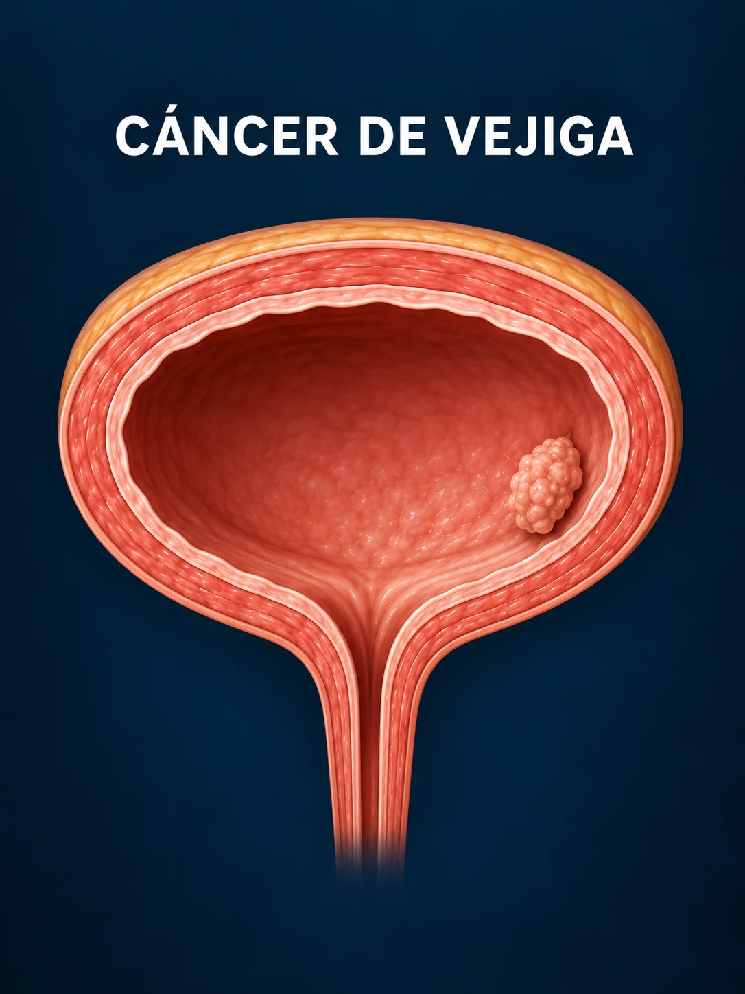
The main symptoms of bladder cancer can vary depending on the stage of the disease and the type of cancer, but they generally include:
1. Hematuria (blood in the urine)
The presence of hematuria (blood in the urine) does not, by itself, confirm the existence of bladder cancer, as there are many other causes of bleeding, such as a urinary tract infection. However, whenever hematuria occurs, it is necessary to rule out bladder cancer (due to its potential severity) by performing tests to determine whether the diagnosis is bladder cancer or another condition.
2. Burning or stinging sensation when urinating
3. Frequent need to urinate
4. Any other type of problems or changes in voiding dynamics
5. In advanced stages or when bladder cancer has spread to other organs, other symptoms may appear, such as:
If you experience any of these symptoms, it is advisable to consult your doctor or urologist.
Age, smoking, and exposure to industrial paints and dyes are the main causes of bladder cancer. In addition, patients with chronic catheterization, recurrent urinary tract infections, or bladder stones may have an increased incidence of squamous cell bladder cancer due to the presence of a foreign body in the bladder.
Bladder cancer is more common in people over 50 or 60 years old, although there are also cases in younger people.
It is the cause of 50% of bladder cancer cases, in both men and women. The probability of a smoker developing bladder cancer is at least three times higher than for non-smokers.
Certain industrial chemicals are linked to bladder cancer, such as aromatic amines used in some dye industries. People exposed to the manufacture of rubber, leather, textiles, paints, and dyes have a higher risk of developing this cancer.
Thus, if both situations are combined, being a smoker and being exposed to this type of substance at work, the risk of suffering from bladder cancer is especially high.
Urinary tract infections, kidney and bladder stones, long-term use of bladder catheters, and other causes of chronic bladder irritation have been associated with squamous subtype bladder cancer.
An infection with a parasite that is ingested through the consumption of untreated water and that lodges in the bladder, also represents a risk factor for squamous subtype bladder cancer, mainly in countries where this parasite is common: Africa and the Middle East, above all.
Prolonged use of the chemotherapy drug cyclophosphamide can irritate the bladder and increase the risk of bladder cancer. People taking this medication are advised to drink plenty of water to help protect the bladder from irritation.
People who receive radiation treatments to the pelvis are more likely to develop bladder cancer.
Although the reasons are not fully understood, white people are more likely to develop bladder cancer than black people.
People with a family history of bladder cancer have a higher risk of developing the disease. They may share changes in certain genes (such as GST and NAT) that impair the breakdown of certain toxins.
On the other hand, a small number of people inherit a genetic syndrome that increases their risk of bladder cancer. This can include:
Several complementary tests are performed for the diagnosis and monitoring of bladder cancer:
It consists of the anatomopathological evaluation of bladder cells that are shed in the urine.
Such as ultrasounds, scans or magnetic resonance imaging.
This involves inserting a camera (cystoscope) through the urethra for direct visualization of the inside of the bladder. This latter procedure is more invasive and uncomfortable for the patient.
Alternatively, we offer a non-invasive test: the Bladder Epicheck® DNA methylation test , which reduces the need for invasive procedures such as cystoscopies. This test is convenient for the patient, requiring only a urine sample.
In the diagnostic process, we ask the patient if they have any risk factors for bladder cancer such as smoking or exposure to chemical agents.
If a tumor is found, we must observe its size, number, location, appearance, level of invasion, and cell grade. Furthermore, this histopathological evaluation determines whether the tumor is non-muscle-invasive , meaning it has not reached the detrusor muscle of the bladder, or whether it is muscle-invasive , meaning its roots have reached the muscular layer (25%). Depending on this, one treatment or another will be applied, allowing us to preserve the bladder in almost all cases of non-muscle-invasive tumors, or requiring a cystectomy (removal of the bladder) in most cases of muscle-invasive tumors (including systemic treatments, such as chemotherapy or immunotherapy, in cases of muscle-invasive tumors).
A CT urogram is also performed to evaluate the ureters, renal pelvis, and calyces and rule out the presence of tumor implants in these locations, since upper urinary tract tumors can coexist with bladder cancer in up to 5-10% of cases. A CT urogram is a radiological procedure that produces three-dimensional cross-sectional images of the human body. As with other imaging tests, an intravenous contrast agent is used. This helps to better delineate anatomical structures, such as the body's blood vessels. Additionally, a thoracoabdominopelvic CT scan is used to rule out the presence of metastases.
In certain cases, the need for an MRI may also arise, especially to assess local invasion in some tumors, or if the patient has a contraindication for a CT scan.
When the tumor is found in an early or superficial stage, it can be treated with a transurethral resection of the bladder (TURB). This surgical procedure is used both for diagnosing bladder cancer and for treating the tumor.
To perform this procedure, a transurethral resectoscope is inserted into the bladder through the urethra (endoscopic procedure). Once inside, a visual examination is performed to identify any tumorous or suspicious lesions (cystoscopy). Subsequently, the tumor tissue is removed (resection) using electrical energy that heats the resectoscope, reaching the optimal temperature to cut and cauterize the affected tissue. In certain cases, the use of laser energy may be indicated instead of electrical energy.
This procedure also allows obtaining a tumor sample for anatomopathological diagnosis and subsequent staging.
This treatment generally requires spinal anesthesia (regional anesthesia injected into the back), although this may vary depending on the patient's needs and the anesthesiologist administering it. The patient must also receive prophylactic antibiotics during surgery to reduce the risk of infection.
This technique ends with the placement of a bladder catheter with a continuous saline flushing system to prevent clot formation.
The duration of this surgery can vary from 30 to 120 minutes depending on the number, size, or location of the tumors. Hospitalization time may last from 1 to 2 days.
Chemotherapy is the use of certain medications to prevent cancer cells from growing or to cause them to die.
In intravesical chemotherapy, drugs are administered directly into the bladder through a catheter inserted into the urethra. It is a local treatment and therefore only destroys superficial tumor cells that come into contact with the chemotherapy solution. It does not reach tumor cells that have infiltrated the bladder muscle or those that have spread to other organs. For this reason, intravesical chemotherapy is only used for non-muscle-invasive bladder cancers. The drugs most frequently used in intravesical chemotherapy are mitomycin-C, gemcitabine, and epirubicin.
Intravesical chemotherapy is administered using different treatment regimens, beginning a few weeks after transurethral resection of the bladder (TURB) to reduce the likelihood of tumor recurrence. There is also the option of administering a postoperative dose of mitomycin C within the first 24 hours after TURB, with the aim of eliminating any tumor cells that may have remained in the bladder after the procedure and thus preventing their implantation in the bladder walls, which could lead to tumor recurrence.
Immunotherapy is the use of drugs that help the immune system recognize and destroy cancer cells and prevent cancer from reproducing and progressing.
Sometimes, after transurethral resection of the bladder, immunotherapy with Bacillus Calmette-Guérin (BCG), a weakened bacterium, may be administered. This can help trigger an immune response when bladder cancer is non-invasive and high-grade. It is administered directly into the bladder via a catheter to prevent tumor recurrence or progression of previous tumors to more advanced stages.
To determine if the patient requires these therapies, several factors are taken into account: local stage, tumor grade, size of the removed lesions, history of previous recurrences or presence of associated carcinoma in situ, as well as the patient's general condition and an evaluation of the other pathologies they present.
Typically, BCG instillations are performed once a week for six weeks during the induction period, and then several boosters are given in three-week intervals with varying frequency for a minimum of one year and a maximum of three years (maintenance period).
First, a transurethral resection of the bladder (TURB) will be performed to establish a diagnosis of tumor infiltration into the muscular layer. Once the diagnosis is made, a radical cystectomy (removal of the bladder) can be performed with curative intent. Cystectomy in men includes the removal of the bladder, prostate gland, and seminal vesicles. In women, it involves the removal of the bladder, uterus, ovaries, cervix, and part of the vagina. Pelvic lymph nodes must also be removed in both sexes.
In the same surgery, the surgeon must create a urinary diversion, that is, a new way to store urine and allow it to be eliminated from the body. There are different techniques for this, which the doctor must evaluate. Among the most commonly used are the neobladder , which involves creating a new bladder from the intestine; the cutaneous ureterostomy , which involves bringing the ureters to the skin of the abdomen; and the cutaneous ureteroileostomy , which involves anastomosing (joining) the ureters to a portion of the intestine, allowing urine to exit through a stoma created in the patient's skin. This surgery can be performed via an open approach; laparoscopically, using a camera that transmits a magnified image to a monitor, allowing the surgeon to operate inside the patient without making large incisions; or robotically , which allows for complex procedures to be performed less invasively and with greater precision, flexibility, and control.
Although cystectomy is usually performed to treat invasive bladder cancer, it can also be performed when the tumor does not invade the muscle but fails to respond to other treatments.
In highly selected patients with invasive cancer, tumors with favorable conditions, or those unsuitable for cystectomy, it is possible to preserve the bladder by treating the patient with a combined treatment of transurethral resection of the bladder (TURB), chemotherapy, and radiotherapy. This is what we call trimodal treatment, and before proceeding, a very thorough evaluation of the tumor and patient characteristics must be carried out by a multidisciplinary team.
This involves administering chemotherapy drugs intravenously. The drugs enter the bloodstream and circulate throughout the body, attacking cancer cells distant from the main tumor—that is, those that have already spread beyond the bladder. This is called systemic chemotherapy.
The most commonly used are cisplatin, gemcitabine, or MVAC, which combines methotrexate, vinblastine, doxorubicin, and cisplatin.
It can also be used after cystectomy in patients with tumor involvement of the perivesical fat or adjacent organs, lymph node involvement, or in patients who already have metastases at the time of diagnosis and have not undergone surgery. Immunotherapy, that is, the use of drugs that help the immune system recognize and destroy cancer cells, is used as a second-line treatment once the disease progresses despite chemotherapy, although there is increasing evidence supporting its early use.
In fact, in the post-cystectomy setting, some patients may be candidates for immunotherapy. On the other hand, the most recent data support the use of chemotherapy plus immunotherapy before cystectomy, followed by immunotherapy after surgery.
The side effects of chemotherapy depend on the individual and the dose used, but can include fatigue, increased risk of infection, nausea and vomiting, hair loss, loss of appetite, and diarrhea. These side effects generally disappear after treatment ends. Immunotherapy has a more favorable toxicity profile than chemotherapy.
Radiation therapy uses intensity-modulated rays as part of multimodal treatment to destroy cancer cells that were not detected during surgery or as palliative treatment for uncontrollable hematuria (bleeding that cannot be contained).
The combination of radiotherapy and chemotherapy after transurethral bladder surgery can sometimes eradicate cancer without the need for cystectomy.
We treat our patients at the ROC Chamberí clinic and in the HM Hospitals network of centers in Madrid: Sanchinarro, Montepríncipe, Madrid Río, Rivas, Puerta del Sur, Nuevo Belén, Policlínico HM Moraleja and Nuevo Norte.

Coming soon
We treat our patients at the ROC Chamberí clinic and in the HM Hospitals network of centers in Madrid: Sanchinarro, Montepríncipe, Madrid Río, Rivas, Puerta del Sur, Nuevo Belén, Policlínico HM Moraleja and Nuevo Norte.
Coming soon

We left very happy with the doctor-patient relationship
JG August 6, 2026
Our patients' trust reflects our commitment to providing excellent, personalized, and attentive urological care. Every experience motivates us to continue improving and offering the care and support each patient needs.


Excellent urological clinic due to the professionals who work there.
A highly professional clinic, with a very high scientific level and advanced technology. They helped me solve my urological problem.

I can only say one thing, the urology girls are the best you can find in this...
I want to express my gratitude to the Roc team, especially Dr. David Carracedo, for their professionalism, the...

Excellent care, both from Dr. Rojo and Dr. Espinales on a medical level and from the staff of...

Top 100 deal

I've been coming to the Urology department at HM Monteprincipe for a long time and I'm very happy with the care I receive from...
Holding in your urine occasionally isn't usually harmful, but when this habit is repeated it can affect bladder function...
Frequent urination in children is often a cause for concern for families, although in many cases it is due to temporary functional changes. Understanding its causes...
In urology, many clinical decisions are based on objective data. It is not enough to know that a patient "urinates worse" or that the stream "has lost..."


Search
Type a keyword and press Enter.