Kidney Stones

This condition can go unnoticed until the stones move through the urinary tract and cause pain.

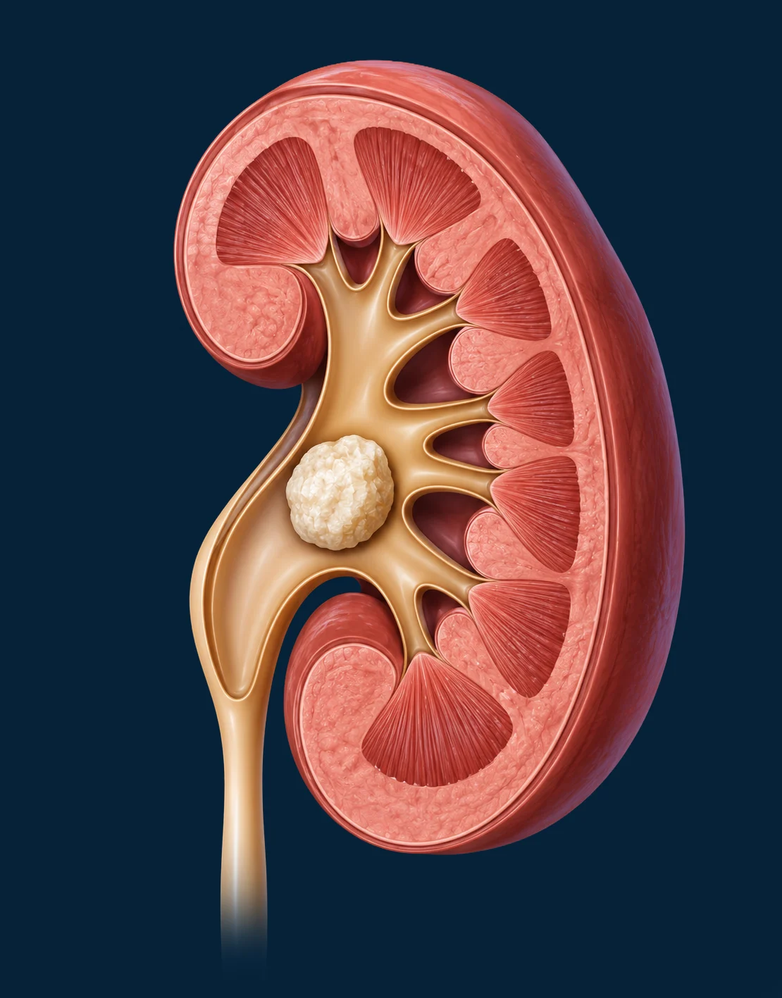
Kidney stones, also known as renal lithiasis, are a very common urological condition. The stones can vary in size, composition, and location, factors that influence their progression and treatment.

( * Required fields)

What is it?

Most people with kidney stones are asymptomatic. Therefore, they are unaware they have them until the stones move within the kidney or pass out—causing the well-known renal colic.

The typical pain associated with kidney stones is renal colic. This pain, which everyone has heard of, begins suddenly in the lower back, radiates to the genital or abdominal area, and may be accompanied by nausea or vomiting. It occurs when a stone becomes lodged, obstructing the flow of urine from the kidney. It is undoubtedly the primary symptom of a kidney stone. In fact, it doesn't necessarily have to be a kidney stone; it can be located elsewhere, such as in the ureter, which is more common. The pain of renal colic is usually relieved or lessened by applying heat or taking warm baths. Unlike lower back pain, backache, or muscle strain, the pain of renal colic is not alleviated by changing position.

Among the symptoms produced by kidney stones, the following may also appear:

  • Fever, due to an associated infection.
  • Nausea.
  • Vomiting
  • Sweating, associated with nausea.
  • Hematuria (blood in the urine), due to the movement of the stone.

Essentially, 85-90% of kidney stones are linked to diet and the lifestyle prevalent in the Western world today. People drink little water and consume more other substances that can cause dehydration or are high in sugar, thus promoting stone formation. Diets high in protein and low in fruits and vegetables also contribute to this, creating an environment conducive to lithogenesis, the scientific term for the formation of stones in the urinary tract.

Most kidney stones are caused by metabolic syndrome, which is also associated with hypertension and diabetes. The most common profile that specialists see in this condition is a middle-aged man who is hypertensive, obese, and diabetic.

There is a very specific type of kidney stone, with a very small percentage (less than 1%), associated with cystine, a hereditary disease with an autosomal recessive inheritance pattern. There may be a familial association in patients who have other diseases with this characteristic, such as hyperparathyroidism, MEN syndromes, and other endocrine-metabolic disorders that manifest clinically as kidney stone formation.

Factors that increase the risk of developing kidney stones include:

  • DehydrationNot drinking enough water every day or drinking other substances high in sugar can increase the risk of developing kidney stones.
  • SubsistenceDiets high in protein, sodium, and sugar can promote the formation of some types of kidney stones.
  • ObesityA high body mass index and weight gain can be associated with an increased risk of developing kidney stones.

Kidney stones are diagnosed through a series of tests. Currently, when a person arrives at the Emergency Department with renal colic , accompanied by sharp and intense lower back pain, the standard procedure is to perform blood tests, urine tests, an abdominal X-ray, and , likely, an ultrasound , at least in the acute phase of the colic.

These tests can detect the presence of kidney stones, their size, and their location. If this is insufficient and the patient shows signs of complications or the problem remains undetermined, a CT scan is recommended . Currently, and as indicated in standard clinical practice guidelines, it is generally agreed that this is the most specific test for determining the presence of kidney stones, their location, size, and composition.

In this regard, when a patient is referred to the clinic, that is, outside of an emergency setting, a detailed study is conducted, beginning with a medical history and followed by an evaluation based on imaging and laboratory tests . A wide variety of imaging tests are available when evaluating a patient with urinary stones:

  • Plain abdominal x-ray.
  • Urological ultrasound.
  • Computed axial tomography (CAT).
  • Magnetic Resonance Imaging.

In addition to these, a metabolic study is usually requested, which consists of slightly more specific blood and urine tests, trying to find any pathology related to the hormones that regulate calcium and vitamin D.

The indication for each of these tests must be assessed by the specialist, not only for the correct diagnosis, but also to plan the most appropriate treatment.

The treatment for kidney stones should be individualized, as it depends on the symptoms, size, and severity. If the stone hasn't caused any symptoms or problems, and isn't large enough, treatment may not be necessary. However, when the stone is large enough to cause an obstruction, it should be treated.

To determine the most appropriate and individualized therapeutic option for each patient, it is necessary to consider the size of the stone, its location, its hardness, its nature, the patient's anatomical factors and associated diseases.

Uretrorenoscopy

Ureterorenoscopy (URS) is an endoscopic technique that involves inserting instruments through the urethra into the bladder. Once in the bladder, the instruments are advanced up the ureter (the tube that carries urine from the kidney to the bladder) to identify the stone, fragment it using a laser, and then remove the fragments.

Currently, laser therapy (in its various modalities such as Holmium-YAG, TFL, or μ:YAG) is considered the first-line treatment for ureteral stones, especially those located in the middle and distal thirds. It is one of the most widely used procedures for managing ureteral stones, given its effectiveness and minimal adverse effects.

This procedure requires anesthesia (general or spinal) and a variable hospital stay, usually 24 hours, although it can sometimes be performed on an outpatient basis. Typically, after the procedure, a double-J ureteral stent is placed to prevent colic in the immediate postoperative period.

Retrograde intrarenal surgery (RIRS)

Retrograde intrarenal surgery (RIRS) is a minimally invasive endoscopic procedure used to treat kidney stones. It allows for the treatment of stones of varying sizes inside the kidney using flexible instruments with an integrated high-definition camera and a high-power light source at the tip.

This technique uses a high-precision and high-power fragmentation source, such as the different lasers we have at ROC, which allow fragmentation-pulverization to be performed with excellent results.

In addition, in certain cases, suction sheaths are used during the procedure, allowing for the continuous removal of fragments and dust resulting from lithotripsy. This improves surgical visibility, reduces intrarenal pressure and surgery time, and facilitates more complete removal of debris.

The RIRS is performed under general anesthesia and requires subsequent hospital admission, although it can be done on an outpatient basis in selected cases, always with early discharge within the first 24 hours. After the procedure, it is usually necessary to place a double J ureteral stent to prevent colic in the immediate postoperative period.

Percutaneous surgery

Percutaneous nephrolithotomy (PCNL) is a minimally invasive surgical procedure used to remove large or complex kidney stones. The term "percutaneous" means "through the skin." This technique is used in cases of:

  • Large stones that cannot be expelled on their own (more than 2 cm in size).
  • They cause intense pain or block the flow of urine.
  • They are located in a place that is difficult to reach using other procedures.
  • They have not responded to other treatments, such as shock wave lithotripsy or RIRS.

The procedure is performed under general anesthesia. A small incision is made in the back, and an image-guided puncture (using ultrasound or X-ray) is performed to create a pathway to the kidney. A nephroscope, a thin instrument with a camera and light, is inserted to locate the kidney stone. Once located, the stone is broken up, and the fragments are removed through the nephroscope.

In many cases, an aspiration sheath is used during the procedure to facilitate the continuous removal of fragments, reduce surgical time, and improve visibility during the procedure, contributing to a more efficient and safe extraction.

Placement of a catheter (optional): In some cases, a small catheter may be placed in the kidney to drain urine and prevent fluid buildup while the incision heals.

Post-operative period: After the procedure, a double-J ureteral stent may be placed to drain urine and prevent fluid accumulation while the incision heals, also preventing the onset of colic in the immediate postoperative period. It is also usually necessary to place a drain that exits the kidney (nephrostomy) and requires hospitalization for 48-72 hours to monitor for any complications.

Combined endoscopic intrarenal surgery (ECIRS)

Combined endoscopic intrarenal surgery (ECIRS) is a surgical technique that embodies the highest excellence in the endourological treatment of kidney stones and requires a large amount of high-tech surgical equipment (lasers, rigid and flexible endoscopes, smart operating rooms).

This endourological technique requires a combined antegrade and retrograde approach to the upper urinary tract, as well as the expertise of a team of experienced surgeons. It is a safe, effective, and minimally invasive procedure for treating all types of urolithiasis.

Similar to percutaneous PCNL surgery, this technique also involves a kidney puncture under ultrasound and X-ray guidance. An antegrade channel is created and dilated to insert a tube of variable caliber through which the stones are fragmented and extracted. In this case, a second surgeon is required who, simultaneously and via a retrograde approach, will reach the renal cavities inaccessible through the antegrade channel to complete the stone fragmentation.

Today, it is the preferred technique, as it allows for the treatment of highly complex kidney stones. Its versatility makes it possible to leave the patient stone-free.

This procedure is performed under general anesthesia. Following the intervention, a double-J ureteral stent is usually placed, and depending on the size of the percutaneous access, a nephrostomy tube may also be created. Hospitalization for 48–72 hours is required.

Morphocompositional analysis of the calculus, lifestyle habits and dietary recommendations

Once urinary stones have been resolved, and in cases where fragments can be recovered, a morphological analysis of the stone should be performed in a laboratory. This test allows for the study of the composition, structure, and shape of the stones once they have been removed from the body. This information is key to identifying their metabolic or dietary origin and trying to prevent future recurrences.

When stones or lithiasic material are not recovered, the composition of the stones can be evaluated by their radiological characteristics and urinary determinations and the macroscopic appearance evaluated during surgery.

What does morphocompositional analysis contribute?

Through microscopic examination (and, in some cases, complementary techniques such as spectroscopy or X-rays), it is possible to determine: the type of stone (calcium oxalate monohydrate or dihydrate, phosphate, uric acid, cystine, etc.), its internal structure (monolayer, multiple layers, nucleus), its possible relationship with metabolic alterations , infections or eating habits, and whether the origin is genetic, dietary or infectious.

Why is the morphocompositional analysis of calculus important?

The information obtained allows the urologist to personalize preventive treatment, correct dietary habits or pharmacological treatments, carry out more effective follow-up and avoid the formation of new stones , which is one of the great challenges in patients with recurrent lithiasis.

This type of analysis is integrated within the personalized medicine approach in urology, which seeks not only to treat, but also to understand why stones form and what measures can be taken to reduce their recurrence.

Recommendations to follow to reduce the risk of developing kidney stones:

  • Increase fluid intake.
  • Reduce the amount of animal protein and salt.
  • Reduce your intake of foods high in oxalates (rhubarb, beets, okra, spinach, Swiss chard, sweet potatoes, nuts, tea, chocolate, black pepper, and soy products).

FAQ

A healthy, varied, Mediterranean diet should be followed, rich in water, avoiding salt, not only the added salt, but also the salt that comes from some foods such as cold cuts, also avoiding sugars and reducing the intake of saturated fats.
Many studies have been conducted on this topic. The findings suggest that if the tap water you drink is of good quality, with low calcium content, there shouldn't be a problem with kidney stone formation. If you're drinking bottled water from the supermarket, it's important to check the label and choose the one with the lowest sodium concentration.
Kidney failure can occur in patients with kidney stones that cause few or no symptoms, as they grow very silently and become large.
A person with kidney stones can and should exercise, as it improves metabolism and is known to promote the elimination of solutes. It's true that physical activity will increase dehydration, especially in summer, requiring more fluid intake than usual to compensate for the fluid lost during exercise.
The postoperative period will depend on the type of procedure performed. If lithotripsy was used, the patient will not require hospitalization, as it is an outpatient procedure. However, if endoscopic surgery was performed, the patient usually remains hospitalized for at least one day, with a double-J stent in place to protect the kidney.
The double J stent has several side effects, such as discomfort and irritation caused by the end of the stent inside the bladder. To minimize these effects, in addition to prescribing medical treatment, the time the stent remains in place is kept to a minimum.
The time a person will need to take a double J catheter It will depend on the complexity of the procedure. Normally, after ureteroscopy or retrograde intrarenal surgery, the recovery time ranges from one week to 10 days. However, if the surgery is more complex, it may be necessary to keep the catheter in place for around 15 days, and this period could be extended to 21 days.
A person with a double J catheter You can lead a more or less normal life to the extent that the catheter allows. It is recommended to drink plenty of water or other fluids and avoid holding your urine for too long. Regarding work, a double J stent does not prevent you from working, as long as the job is not excessively physical. There would also be no problem with exercising, but it could have some associated consequences, such as blood in the urine.

Our centers
urology

We treat our patients at the ROC Chamberí clinic and in the HM Hospitals network of centers in Madrid: Sanchinarro, Montepríncipe, Madrid Río, Rivas, Puerta del Sur, Nuevo Belén, Policlínico HM Moraleja and Nuevo Norte. 

ROC Chamberí

Coming soon

ROC Bernabéu

ROC HM Sanchinarro

ROC HM Montepríncipe

ROC HM South Gate

ROC HM Madrid Río

ROC HM Rivas

ROC HM New Bethlehem

ROC HM New North

ROC Polyclinic HM Moraleja

Our centers
urology

We treat our patients at the ROC Chamberí clinic and in the HM Hospitals network of centers in Madrid: Sanchinarro, Montepríncipe, Madrid Río, Rivas, Puerta del Sur, Nuevo Belén, Policlínico HM Moraleja and Nuevo Norte. 

ROC Clinic

Urology in Madrid

4,8 ★ ★ ★ ★ ★★ ★ ★ ★ ★
make an appointment now
★ ★ ★ ★ ★★ ★ ★ ★ ★ Verified rating

We left very happy with the doctor-patient relationship

JG August 6, 2026

View all ratings (43)
Powered by TOPDOCTORS

The experience of
our patients

Our patients' trust reflects our commitment to providing excellent, personalized, and attentive urological care. Every experience motivates us to continue improving and offering the care and support each patient needs.

4,7
★★★★★ 94 reviews published in Google
Leave us a review on Google

Related articles

27/10/2026
Urological ultrasound: what it is, what it detects and how it is performed

Urological ultrasound is a non-invasive imaging test that allows the study of different organs of the urinary system, mainly the kidneys and bladder, and...

21/05/2026
Changes in urine during pregnancy: what's normal and when to seek medical advice

During pregnancy, changes in urine may occur, such as increased urinary frequency, changes in color, or discomfort when urinating. Learn more...

20/01/2026
Strong-smelling urine: common causes and when it's normal

“Doctor, my urine smells strong. Is this normal?” This is one of the most common questions in urology, and in most cases, the…