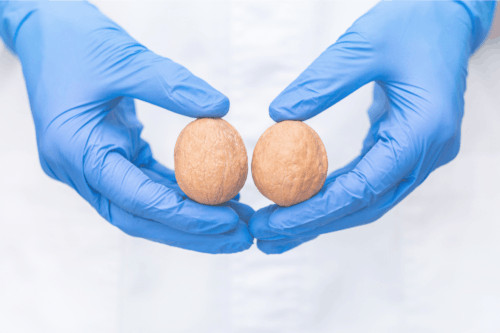
Cryptorchidia

Cryptorchidism or testicular maldescent is a very common anomaly in pediatric boys and is even more common in premature infants. It occurs when the testicle does not reach its normal position in the scrotal sac during pregnancy. It is identified because the scrotal sac on that side is empty. In up to 30% of cases, cryptorchidism can affect both sides.
During the first 6 months of life the testicle can spontaneously descend into the scrotal sac without intervention, unless there is an inguinal hernia associated with cryptorchidism.
If the testicle does not reach its physiological position, it is important that the child be evaluated by a specialist in pediatric surgery, who will explore the inguinal and pelvic region if necessary, to determine the position and characteristics of the testicle and the need or not for surgical treatment. He will also determine the presence or not of associated anomalies, which may require a genetic or endocrine study as a priority.
It is important to emphasize that this consultation should not be delayed, since the testicle needs specific conditions for its normal development that it will not have if it is, for example, in an intra-abdominal location.
Cryptorchid testicles can be divided into palpable and non-palpable, this classification being fundamental to decide the treatment.
Palpable testes include:
- Testicular maldescent: these are testicles that interrupted their way to the scrotal sac and were trapped at some point along the route from the lumbar region, being frequent that they remain in the inguinal region.
- Ectopic testicle: are those testicles that deviated from their path during descent and are located in the femoral, pubic or perineal region. These testicles must be intervened for their correct position.
- Retractile testis: these are testicles that have reached their normal position in the scrotal pouch, but due to an exacerbated cremasteric reflex, they are introduced into the abdominal cavity during physical examination. These testicles remain in the pouch without tension but should be monitored by a pediatric specialist since, in up to one third of cases, they may ascend and not return to their correct position.
- Trapped testicle: it is the testicle that was trapped in the scar of a surgical intervention in the inguinal region.
The non-palpable testes are:
- Intra-abdominal or canalicular testis: up to 50-60% of cases. Sometimes they are close to the deep inguinal orifice and can come out of the abdominal cavity into the inguinal canal. These testicles do not descend on their own and must be operated to promote their correct development.
- Agenesis testicle: up to 4% of cases. It occurs when one of the testicles did not form or atrophied during pregnancy.
Diagnosis of cryptorchid disease
In the diagnosis of cryptorchidism, the clinical history is very important. It is important to know the maternal history of medication consumption (corticosteroids), possible pregnancy problems, family history of unexplained neonatal deaths, genital anomalies, etc.
An examination by a pediatric surgeon will also be performed to rule out possible associated anomalies and to make sure that the cremasteric reflex, so exacerbated in some children, does not mask the true location of the testicle.

Imaging studies such as ultrasound are not usually necessary, as ultrasound has limited sensitivity in identifying ectopic testes, but may be indicated in some patients to identify associated abnormalities.

Cryptorchid treatment
Currently, the treatment of cryptorchidism is surgical and is performed from 12-18 months of age so that the testicle can develop normally, since the chances of spontaneous descent after that age are very low.
Depending on the location and whether or not it is a palpable testicle, a laparoscopic approach or an inguinal incision will be chosen .
- Palpable testicles are approached via the inguinal approach to close the communication with the abdominal cavity and fix the testicle in the scrotal pouch. This is generally an outpatient procedure in which the patient goes home a few hours after surgery.
- In patients operated by laparoscopic approach, the surgeon will identify the testicle and the possibility of descending it into the scrotum in a single procedure or if, on the contrary, it is necessary to do it in two stages in case the vessels that bring irrigation to the testicle are very short and do not allow it.
During this procedure, the surgeon makes four incisions, one to insert the camera, two other working trocars and a scrotal incision to fix the teste in the pouch.
It is possible that the hospital stay may be slightly longer for this type of intervention, but rarely more than a day.
Team of the Cryptorchid Unit


 +34 912 627 104
 Contact


