Diagnosis Kidney Cancer
In recent years, thanks to imaging tests, the number of diagnoses in early stages has increased.
Kidney cancer usually does not produce symptoms until more advanced stages. For this reason, more than half of the cases are diagnosed incidentally in diagnostic tests performed for another reason.
- Super-specialized urologists
- Personalized treatment
- Minimally invasive approach
- More than 16,000 patients successfully treated
Diagnosis of kidney cancer
More than half of kidney tumors are diagnosed incidentally in the course of diagnostic tests performed for another reason, when the tumor has not yet caused any symptoms. If this has not occurred, but there is suspicion of kidney cancer, a series of diagnostic tests will be carried out:
- Assessment of complete medical history.
- Laboratory analysis of urine to check for blood or cancer cells.
- Blood tests, which will reveal whether the kidneys are functioning properly.
- Complete blood count: measures the number of blood cells in the blood, such as white blood cells, red blood cells and platelets. People with kidney cancer may have anemia - low red blood cell counts.
- Chest x-ray: performed to check if the cancer has spread to the lungs.
- CT: should be performed in all cases to characterize the renal mass, evaluate neighboring organs and distant extension. It is a special type of radiography that captures detailed images to know if the cancer has spread.
- Magnetic resonance imaging (MRI): It is used in cases of diagnostic doubt, in cases of complex cystic lesions, if the patient is allergic to iodinated contrasts or to evaluate the possible infiltration of the renal and cava vein. This test uses radio waves and magnets, instead of X-rays, to show the soft tissue parts of the body. It will reveal whether the cancer has spread.
- Ultrasound: uses sound waves to produce images of the inside of the body to diagnose solid kidney masses and differentiate them from cystic lesions. If a biopsy is needed, ultrasound can guide the needle into the mass to extract cells for analysis.
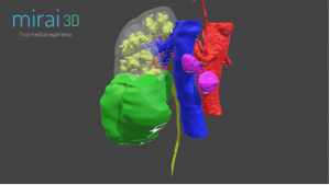
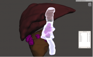
- 3D models: customized 3D models are of utmost importance in the surgical planning of renal cancer, regardless of its extent. They provide very detailed and accurate information on the anatomy of the tumor, its relationships with the rest of the kidney and neighboring viscera, the anatomy of the affected kidney and its vascular supply. These 3D models can be made available virtually or physically thanks to 3D printers. Whether the planned surgery is conservative or radical, they provide real-time, highly accurate information to the surgeon who will direct the intervention, who can navigate the anatomical details of each individual case in order to provide a tailor-made treatment.


- Biopsy: Ultrasound or CT guided biopsy of the renal mass is only advised in selected cases. For this test, a small amount of tissue is removed to reveal whether cancer cells are present. For most cancers, this is the only way to determine the disease with certainty. In the case of kidney cancer, this test is not always necessary, as x-rays and other imaging tests may be sufficient. For the biopsy of a kidney tumor, one or more samples of the tumor are taken with a fine needle under local anesthesia. This procedure may cause blood in the urine (hematuria). Rarely, more serious bleeding may occur, although this is usually a low-risk procedure.
The imaging tests described above provide essential information on the size of the tumor, its extent, its potential invasion of local veins such as the inferior vena cava, the status of lymph nodes or neighboring organs. This is important in determining further treatment.
With the results of these tests and their individualized diagnosis, the urologist will be able to define the stage of the disease. By analyzing the tumor tissue, received during surgery or biopsy, the pathologist determines the subtype of the tumor and whether or not it is aggressive. Together, the stage, subtype and aggressiveness of the tumor form the classification.
There are a number of poor prognostic factors that can help us predict the course of the disease: local or distant extension (metastases), high nuclear histologic grade (Furhman grade), onset of symptoms, anemia, high erythrocyte sedimentation rate (ESR), elevated alkaline phosphatase or elevated lactate dehydrogenase (LDH).
Assignment of kidney cancer grade
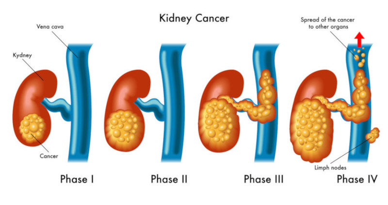
Renal tumor staging is used to estimate your individual prognosis. Based on this individualized prognosis, your doctor will discuss the best course of treatment for you. Kidney cancers are generally assigned a grade from 1 to 4.

- Stage I: the cancer cells are very similar to normal kidney cells and only affect this organ.
- Stage II: The cancer invades the capsule surrounding the kidney.
- Stage III: The cancer invades the vena cava or renal vein or affects neighboring lymph nodes.
- Stage IV: cancer cells are very different from normal cells and tend to grow faster. The cancer invades neighboring organs or produces distant metastases.
They ask us in the Consultation
How do you know if a cyst in the kidney is malignant?
To know if a kidney cyst is malignant, it is important to perform a series of studies and medical evaluations. The most common steps to determine the nature of a kidney cyst are: Renal ultrasound: ultrasound can show the shape and size of the cyst, as well as the presence of any suspicious features, such as irregular walls or inhomogeneous fluid. Computed tomography (CT): If the cyst has suspicious features on ultrasound, a CT scan may be done to obtain more detailed images. This helps to identify if there are solid masses within the cyst or changes in the cyst walls that suggest malignancy. Magnetic resonance imaging (MRI): is useful to better characterize the cyst and may provide additional information about its composition. This is particularly useful if there is doubt after the CT scan. Bosniak classification: The Bosniak classification system is used to categorize renal cysts based on their appearance on imaging and help determine the risk of malignancy. Biopsy: In some cases, if the cyst is suspected to be malignant or if imaging tests are inconclusive, a renal biopsy may be performed to obtain a sample of the tissue and examine it under the microscope. In general, benign kidney cysts do not cause symptoms. However, if a cyst becomes large, it may cause pain in the back or abdomen. If the cyst is malignant, there may be additional symptoms such as blood in the urine, unexplained weight loss or fatigue.
What are the symptoms of advanced kidney cancer?
Kidney cancer does not present symptoms until there is tumor growth. When tumor growth has already occurred, symptoms usually include pain, appearance of an abdominal mass or blood in the urine.
What are the causes of kidney cancer?
It is usually related to smoking and obesity. Although there are also other risk factors such as age or first-degree family history that may play a role.
Where does kidney cancer metastasize?
The most frequent areas to which kidney cancer can spread are the bones, liver, lungs, brain and distant lymph nodes.
Team of the Kidney Cancer Unit
Newsof ROC Clinic in Kidney Cancer
Research
Da Vinci and Hugo RAS Platforms for robot-assisted partial nephrectomy: a preliminary prospective comparative analysis of the outcomes.


 +34 912 627 104
 Contact


